Copyright© KANEHARA & Co., LTD. All Rights Reserved.
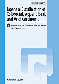
大腸癌取扱い規約 (英語版) 第3版 Japanese Classification of Colorectal,Appendiceal,
『大腸癌取扱い規約第9版』(2018年7月)の全文英訳版。

| 編 集 | 大腸癌研究会 |
|---|---|
| 定 価 | 4,620円 (4,200円+税) |
| 発行日 | 2019/04/25 |
| ISBN | 978-4-307-20395-1 |
B5判・136頁・図数:11枚・カラー図数:52枚
| 在庫状況 | あり |
|---|
癌の分類・記載法として、本邦で広く使用されている癌取扱い規約。諸外国との交流が進む大腸癌診療において、規約をもとに収集されたデータによる研究成果の海外発信や海外の読者による論文の理解のために、英語版の刊行が求められている。日本語版規約第7版補訂版準拠の英語版第2版(2009年)より10年ぶりの改訂となる本版は、『大腸癌取扱い規約第9版』(2018年7月)に準拠した全文英訳版である。
 Purchase this book from overseas
 Purchase this book from overseas
Table of Contents
The principles of revision and changes from the previous edition(the 8th Japanese edition).
I. Guidelines for classification
1 Aims and subjects
1.1 Aims
1.2 Subjects
2 General principles of description methods
2.1 Clinical, surgical, and pathological findings
2.2 Findings following preoperative treatment
2.3 Findings of recurrence
3 Recording of findings
3.1 Primary tumors
3.1.1 Tumor location
3.1.2 Anatomical divisions of colon and rectum, vermiform appendix, and anal canal
3.1.3 Circumferential divisions of the wall of the rectum and anal canal
3.1.4 Number and size of lesions and proportion of the tumor in relation to the circumference of the bowel
3.1.5 Macroscopic types
3.1.5.1 Main macroscopic types
3.1.5.2 Subtypes of macroscopic type 0
3.1.6 Depth of tumor invasion(T)
3.2 Metastasis
3.2.1 Lymph node metastasis
3.2.1.1 Lymph node groups and station numbers
3.2.1.2 Lymph node station number
3.2.1.3 Regional lymph nodes
3.2.1.4 Lymph node metastasis(N)
3.2.2 Distant metastasis(M)
3.2.2.1 Liver metastasis(H)
3.2.2.2 Peritoneal metastasis(P)
3.2.2.3 Pulmonary metastasis(PUL)
3.3 Stage grouping
3.3.1 Clinical and pathological classifications for stage grouping
3.3.2 Stage grouping following preoperative treatment
3.4 Multiple colorectal cancers, multiple primary cancers
3.5 Family history and hereditary diseases
4 Endoscopic and surgical treatments
4.1 Endoscopic treatment
4.1.1 Endoscopic treatment method
4.2 Surgical treatment
4.2.1 Approach to the lesion
4.2.2 Surgical procedures
4.2.3 Lymph node dissection
4.2.3.1 Extent of lymph node dissection(D)
4.2.3.2 Extent of lateral lymph node dissection(LD)
4.2.4 Anastomosis
4.2.4.1 Types of anastomosis
4.2.4.2 Methods of anastomosis
4.2.5 Combined resection of adjacent organs and structures
4.2.6 Preservation of autonomic nerves(AN)
5 Assessment of cancer involvement at resected margin, residual tumor, and curability
5.1 Cancer involvement at resection margins
5.1.1 Specimens obtained by endoscopic resection
5.1.1.1 Horizontal margin(lateral/mucosal margin)(HM)
5.1.1.2 Vertical margin(deep/intramural margin)(VM)
5.1.2 Specimens obtained by surgical resection
5.1.2.1 Proximal margin(PM)
5.1.2.2 Distal margin(DM)
5.1.2.3 Radial margin(circumferential resection margin)(RM)
5.2 Residual tumor
5.2.1 Residual tumor following endoscopic treatment(ER)
5.2.2 Residual tumor following surgical treatment(R)
5.3 Curability
6 Chemotherapy and radiotherapy
6.1 Chemotherapy documentation
6.2 Radiotherapy documentation
6.2.1 Aims of radiotherapy
6.2.2 Methods of radiotherapy
6.2.3 Radiation field
7 Evaluation of resected specimens
7.1 Macroscopic findings
7.1.1 Tumor location
7.1.2 Macroscopic types
7.1.3 Size
7.1.3.1 Tumor size
7.1.3.2 Size of the intramucosal component of the tumor
7.1.3.3 Size of the ulcerated area
7.1.4 Proportion of the tumor in relation to the circumference of the bowel
7.1.5 Distance from the lesion to the resection margin
7.1.6 Extent and properties of invasion and metastasis
7.1.7 Depth of tumor invasion
7.1.8 Lymph node metastasis and location
7.2 Histological findings
7.2.1 Histological types
A. Colon and rectum
B. Vermiform appendix
C. Anal canal(including the perianal skin)
7.2.2 Infiltration pattern(INF)
7.2.3 Lymphovascular invasion
7.2.3.1 Lymphatic invasion(Ly)
7.2.3.2 Venous invasion(V)
7.2.4 Tumor budding(BD)
7.2.5 Extramural cancer deposits without lymph node structure(EX)
7.2.6 Perineural invasion(Pn)
7.3 Histological criteria for the assessment of response to chemotherapy/rediotherapy
7.4 Histological assessment of biopsy specimens(Group classification)
7.5 Measurement of the depth of invasion
7.5.1 T1 cancer
7.5.2 Tumor invasion beyond the MP in sites without the serosa
8 Treatment outcome record
8.1 Number of patients
8.2 Multiple colorectal cancers, multiple primary cancers
8.3 Modalities of treatment and adjuvant therapy
8.4 Total number of colorectal cancer cases with treatment, and the number and rate of cases by treatment types
8.4.1 Resection rate
8.4.2 Endoscopic treatment
8.4.3 Chemotherapy and radiotherapy
8.5 Number and rate of operative mortality
8.6 Number and rate of hospital mortality following surgery
8.7 Survival analysis
8.7.1 Survival
8.7.2 Recurrence/metastasis; site(s)and mode
8.7.3 Survival analysis method
Supplement: Lymph node groups and station number
Supplementary Reference of Macroscopic Types
Supplement: Measurement of submucosal invasion distance
II. Assessment of response to chemotherapy and radiotherapy
1 Assessment of response
2 Definition of efficacy endpoints
2.1 Response rate
2.2 Overall survival(OS), progression-free survival(PFS), relapse-free survival(RFS), disease-free survival(DFS), time to treatment failure(TTF)
3 Documentation of adverse events
III. Explanation of pathological items[Supplement: Histology Atlas]
1 Histological types
A. Colon and rectum
B. Vermiform appendix
C. Anal canal(including the perianal skin)
2 Histological assessment of biopsy specimens(Group classification)
3 Handling of resected specimens
3.1 Handling of biopsy materials
3.2 Macroscopic examination and handling of surgically resected specimens
3.3 Handling of endoscopically resected specimens
Supplementary histology atlas
Supplements
Supplement 1 TNM Classification of malignant tumours
Supplement-1-1 Carcinoma of the colon and rectum
Supplement 1-2 Carcinoma of the appendix
Supplement 1-3 Carcinoma of the anal canal
Supplement 1-4 Well-differentiated neuroendocrine tumours(G1 and G2)of the colon and rectum, and the appendix
Supplement 2 Summary of findings
Supplement 3 Checklist for pathological report
Supplement 4 List of abbreviations
Index
Preface of the Third English Edition
The Japanese Society for Cancer of the Colon and Rectum(JSCCR)is pleased to publish the third English edition of the Japanese Classification of Colorectal, Appendiceal and Anal Carcinoma(JCCRC), which is the translated version of the ninth Japanese edition of the JCCRC published in July 2018. The previous English edition published in January 2009 is the translated version of the seventh Japanese edition of JCCRC.
Our classification and treatment strategies for colorectal cancer, which have been developed based on accumulating clinical and histopathological studies on colorectal cancer in patients treated during a span of 45 years in Japan, differ from those implemented in Western countries in certain regards. Similar to trends observed in industry, economy, society, and culture, among others, medical care is also affected by globalization. The latest edition of the JCCRC is focused on harmonization with the TNM classification. However, several differences still exist, which stem from unique and detailed surgical approaches and histopathological studies, such as those involving main lymph nodes around the feeding arteries, lateral lymph nodes on the pelvic side wall for rectal cancer, and extramural discontinuous cancer spread and tumor budding. We believe that these are important aspects for a more precise evaluation of the spread in colorectal cancer and a more proper estimation of prognosis.
Furthermore, strategies and recommendations for treatment of colorectal cancer, which were included in the JCCRC before 2005, when the first edition of the Japanese guidelines for treatment of colorectal cancer was published, have been moved to the guidelines for the JCCRC because the Japanese guidelines for colorectal cancer have been improved through several editions since 2005.
The number of clinical and histopathological studies on colorectal cancer conducted in Japan and published in English medical journals has been increasing significantly. Most of these studies rely on data collected according to the JCCRC. Publication of the English version of the recently revised JCCRC will contribute immensely toward a better understanding of these articles by non-Japanese readers.
February 2019
Kenichi Sugihara
President,
Japanese Society for Cancer of the Colon and Rectum
