Copyright© KANEHARA & Co., LTD. All Rights Reserved.
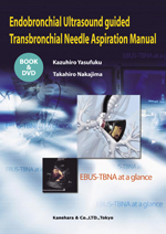
超音波気管支鏡ガイド下針生検マニュアル 英語版
「超音波気管支鏡ガイド下針生検マニュアル」の英訳版。

| 著 者 | 安福 和弘 / 中島 崇裕 |
|---|---|
| 定 価 | 8,800円 (8,000円+税) |
| 発行日 | 2009/08/19 |
| ISBN | 978-4-307-20269-5 |
B5判・96頁・カラー図数:246枚
| 在庫状況 | なし |
|---|
Endobronchial ultrasound-guided transbronchial needle aspiration(EBUS-TBNA)using the convex-probe endobronchial ultrasound(CP-EBUS)has emerged as a new minimally invasive technology for the evaluation of the mediastinum as well as the hilum. The main purpose of the development of the CP-EBUS was for the use in lymph node staging of lung cancer. We began the development of this device in collaboration with Olympus Corporation in early 2002. Preliminary studies looking at animal models enabled us to move to clinical trials. It has now become the standard of mediastinal staging in lung cancer patients. EBUS-TBNA is now being applied to other diseases in the Thorax other than lung cancer. Although there is a significant demand for this procedure, it is still not widely available and the procedure is not routinely taught.
EBUS-TBNA requires the skill of performing a regular bronchoscopy as well as knowledge of technical aspects of the procedure. It allows bronchoscopists to see beyond the airway, thus understanding of the mediastinal and hilar anatomy is critical for a successful and safe procedure. This book explains in details what a bronchoscopist needs to know for performing a successful EBUS-TBNA. The principle of endobronchial ultrasound, preparation of the device, a step by step guide for the procedure as well as techniques for the handling of the specimen obtained by EBUS-TBNA is described. In addition, we have included clinical cases diagnosed by EBUS-TBNA for reference. The anatomy of the mediastinum and the hilum from a bronchoscopic view will guide bronchoscopist to a successful EBUS−TBNA. The DVD included in this book demonstrates how the authors perform EBUS-TBNA emphasizing technical aspect of the procedure. This will ensure readers to fully understand how EBUS-TBNA should be performed.
EBUS-TBNA requires the skill of performing a regular bronchoscopy as well as knowledge of technical aspects of the procedure. It allows bronchoscopists to see beyond the airway, thus understanding of the mediastinal and hilar anatomy is critical for a successful and safe procedure. This book explains in details what a bronchoscopist needs to know for performing a successful EBUS-TBNA. The principle of endobronchial ultrasound, preparation of the device, a step by step guide for the procedure as well as techniques for the handling of the specimen obtained by EBUS-TBNA is described. In addition, we have included clinical cases diagnosed by EBUS-TBNA for reference. The anatomy of the mediastinum and the hilum from a bronchoscopic view will guide bronchoscopist to a successful EBUS−TBNA. The DVD included in this book demonstrates how the authors perform EBUS-TBNA emphasizing technical aspect of the procedure. This will ensure readers to fully understand how EBUS-TBNA should be performed.
Chapter1 Principles and Practice of Endobronchial Ultrasound guided Transbronchial Needle Aspiration
I.General Statement
1.Introduction
2.The clinical application of EBUS-TBNA
3.The limitation of EBUS-TBNA
II.Basics of Ultrasound
1.Principles of Ultrasound
2.Basic Imaging Modes
3.Scanning Methods
4.Resolution
5.Artifacts
III.Instrument
1.Convex Probe Endobronchial Ultrasound(CP-EBUS, BF-UC160F-OL8)
2.Ultrasound Processor(EU-C60/EU-C2000)
3.Dedicated EBUS-TBNA needle(NA-201SX-4022)
IV.Preoperative Preparation
1.Instrumental Preparation
2.Preparation of the Needle
3.Anesthesia
4.Training for EBUS-TBNA
V.Procedural Technique
1.Insertion of the Bronchoscope
2.Visualization of Lymph Nodes
3.EBUS-TBNA
VI.Processing the Specimen
Chapter2 Bronchoscopic and EBUS Anatomy of the Mediastinum and the Hilum
I.Bronchoscopic Anatomy of the Mediastinum and the Hilum
1.Lower Trachea and Carina
2.Right Main Bronchus
3.Right Middle and Lower Lobe Bronchi
4.Upper Trachea
5.Left Main Bronchus
6.Left Upper and Lower Lobe Bronchi
II.Systematic Assessment of Mediastinal and Hilar Lymph Nodes
1.Right Hilar Lymph Nodes to Subcarinal Lymph Nodes
2.Left Hilar Lymph Nodes to Subcarinal Lymph Nodes
3.Superior Mediastinal Lymph Nodes
Chapter3 Clinical Cases
I.Primary Lung Cancer
1.Lymph Node Staging in Lung Cancer
2.Comparison of PET, CT and EBUS-TBNA for Lymph Node Staging
3.Restaging of the Mediastinum by EBUS-TBNA
4.Evaluation of post-operative Mediastinal Lymphadenopathy in Lung Cancer Patients
II.Metastatic Lung Tumors
◆Case1 Pulmonary metastasis from colon cancer that diagnosed by EBUS-TBNA of mediastinal/hilar lymph nodes
◆Case2 A case of pulmonary metastasis from breast cancer with mediastinal/hilar lymph node metastasis diagnosed by EBUS-TBNA
◆Case3 Lung metastasis from thyroid cancer diagnosed by EBUS-TBNA
◆Case4 Pulmonary metastasis from renal cell cancer with mediastinal/hilar lymph node involvement diagnosed by EBUS-TBNA
◆Case5 Evaluation of chemotherapy by EBUS-TBNA in pulmonary metastasis from germ cell tumor
◆Case6 A case with query mediastinal/hilar lymph node metastasis after surgery for esophageal cancer and pulmonary cancer
III.Diagnosis of Intrapulmonary Tumors
◆Case1 A case of lung cancer adjacent to the trachea diagnosed by EBUS-TBNA
◆Case2 A case of squamous cell carcinoma in the superior segment of the right lower lobe diagnosed by EBUS-TBNA
◆Case3 A case of a pulmonary metastasis from colon cancer near the lobar bronchus of the left lung diagnosed by EBUS-TBNA
IV.Mediastinal Tumors
◆Case1 Invasive thymoma with mediastinal lymph node metastasis diagnosed by EBUS-TBNA
◆Case2 Treatment of mediastinal cyst by EBUS-TBNA
◆Case3 Spinal chondrosarcoma diagnosed by EBUS-TBNA in a patient with multiple osteochondromatosis
◆Case4 Primary mediastinal goiter diagnosed by EBUS-TBNA
V.Lymphoma
◆Case1 Malignant lymphoma diagnosed by EBUS-TBNA
◆Case2 Castleman’s tumor diagnosed by EBUS-TBNA
VI.Sarcoidosis
◆Case1 Diagnosis of sarcoidosis by EBUS-TBNA following a non-diagnostic transbronchial biopsy
◆Case2 Spinal sarcoidosis diagnosed by EBUS-TBNA
VII.Miscellaneous
◆Case1 Primary amyloidosis involving mediastinal lymph nodes diagnosed by EBUS-TBNA
◆Case2 Mediastinal adenopathy caused by eosinophilic pneumonia
◆Case3 Pulmonary tuberculosis diagnosed by EBUS-TBNA
◆Case4 Assessment for EGFR mutation using EBUS-TBNA sample and administration of Gefitinib
